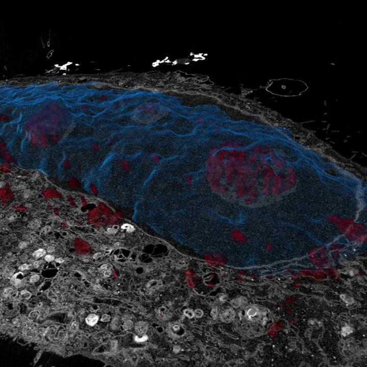

생명체의 초미세 구조로부터의 새로운 발견 - 생명과학 연구의 향상을 위한 Volume EM을 웨비나를 통해 확인하세요!
무료 웨비나 시리즈에 등록하고, 경험하세요.볼륨 전자현미경 (vEM)으로 알려진 SEM의 기술과 방법을 통해 복잡한 초미세구조 정보를 얻을 수 있습니다. 연구 커뮤니티와 상업 커뮤니티 간의 파트너십과 과학적 발전으로 이러한 방법들이 전자현미경에 대한 경험이 거의 없는 사람들에게도 사용하기 쉽고, 접근하기 쉬워졌습니다.
이 웨비나 시리즈에서는 vEM 이미징의 기술적 토대을 살표보고, 계속 발전하고 있는 응용 분야를 강조하여 소개합니다.
먼저 vEM을 위한 샘플 준비를 먼저 살펴보고, 이어서 Array Tomography Serial Block-Face SEM과 FIB-SEM과 같은 개별 vEM 기술을 자세히 살펴볼 예정입니다.
이 웨비나 시리즈는 워크플로 중심의 소프트웨어 솔루션의 발전된 이미지 처리, 데이터 분석 및 결과 시각화 기능에 대한 통찰력까지 다루게 됩니다.
이 6부작으로 구성된 웨비나 시리즈에 참여하여 vEM이 어떻게 신경생물학, 암 연구, 발생학, 식물 과학 등 다양한 분야에서 새로운 발견을 이끌어내는지 알아보세요.
웨비나 발표자


Kirk Czymmek
Principal Investigator, Director, Advanced Bioimaging Laboratory Donald Danforth Plant Science CenterDr. Kirk Czymmek received his Ph.D. in Botany and Plant Pathology and has over 30 years’ experience dedicated to advanced microscopy techniques including most forms of light, x-ray, electron microscopy and correlative microscopy. His work has focused on developing and applying cutting-edge microscopy tools for imaging cells, tissues, and biomaterials.


Christel Genoud, Ph.D
Senior Lecturer, Faculty of Biology and Medicine & CEO of the Dubochet Center for Imaging EPFL and Universities of Lausanne, Geneva, and Bern, SwitzerlandChristel Genoud is an electron microscopist with a background in neuroscience. She studied at the University of Lausanne and got her PhD studying the plasticity of the mouse cortex with electron microscopy under the supervision of Graham Knott. She then went in industry and worked for a company to develop and launch the volumeSEM technique inspired by the microtome developed by W. Denk. In 2008, she joined the Friedrich Miescher Institute in Basel (a Novartis Research Foundation affiliated to the University of Basel) and built an EM facility focuses on volume SEM techniques. From 2016-2020, she managed as well the cryo-EM facility of the FMI shared with Novartis contributing to its creation. Since 2020, she is leading the Em facility of the University of Lausanne. In 2022, she has been nominated CEO of the Dubochet Center for Imaging, a multi-institutional cryo-EM center with platforms in three universities , Lausanne, Geneva and Bern as well as at EPFL.


Corrado Cali, Ph.D.
Associate Professor, Department of Neuroscience University of Turin, ItalyCorrado Calì was trained an electronic engineer at Politecnico di Torino, Italy. He completed his MSc in 2006, in the lab of Henry Markram at EPFL (Lausanne, Switzerland), where he became interested in neuroscience. In the same year he joined the lab of Paola Bezzi at UNIL (University of Lausanne, Lausanne, Switzerland), where he got his doctoral degree in 2012. His research was focused in elucidating the physiological role of astroglial cells in the modulation of synaptic transmission, by releasing neuroactive compounds (so called "gliotranmsission"). After one year as postdoctoral fellow in Graham Knott’s lab, where he enriched and deepend his skills in the state-of-the-art electron microscopy techniques, he joined Pierre Magistretti’s lab in KAUST (King Abdullah University of Science and Technology, Thuwal, Saudi Arabia). Here, he investigated the mechanisms of metabolic support of astrocytes to neurons using morphological and 3D imaging approaches. In particular, he pioneered the use of VR (virtual reality) in neuroscience and is actively exploring scientific visualization approaches for explorative analysis of sparse and dense reconstructions of the CNS from serial-section Electron Microscopy. His interests in 3D visualization and augmented reality led him to establish a company, Intravides (Torino, Italy) with a more clinical focus, developing an Augmented Reality medical device to assist and improve neurosurgical training and practice. Since February 2020, he joined the department of Neuroscience of the University of Torino (Italy), and is now associate professor of Human Anatomy.


Jean Daraspe
Expert Scientist, Deputy Head Universite de LausanneJean obtained his master’s degree in plant sciences at the University of Paris XI in 2005 using confocal microscopy. He acquired his knowledge of electron microscopy after joining the team of Dr. Jean-Marc Verbavatz at the CEA in Saclay, France. Jean joined the EMF in 2008 as a research scientist. He is in charge of all aspects of sample preparation available at the EMF for transmission electron microscopy (TEM) preparation and imaging, using his years of practical experience with biological samples. He is also involved in the development of informatics tools for the automatic image acquisition and analysis of bio-EM samples. Jean trains users in electron microscopy during one-on-one hands-on training sessions and in workshops given by the facility.


Jemima Burden, Ph.D
Head of Electron Microscopy, University College LondonJemima is Head of Electron Microscopy at the Laboratory for Molecular Cell Biology (LMCB) at University College London (UCL). She received her BSc from University of Bath and her PhD from Imperial College London, where her fascination for microscopy began, before specialising in electron microscopy at the LMCB.


Ian White, Ph.D
Deputy Head of Electron Microscopy, LMCBIan is Deputy Head of Electron Microscopy at the LMCB. Having completed his PhD at the University of Leeds and a postdoctoral position at the University of Sheffield, Ian moved to UCL where he began to learn the techniques involved in biological electron microscopy, before joining the LMCB as a specialist.


Laura Matino
Research fellow, School of Medicine and Surgery, Università degli studi di Milano BicoccaLaura Matino received her education in biomedical engineering at Università degli studi di Napoli Federico II where she completed her MSc in 2017. She then joined the Tissue-electronics research group at the Center of Advanced Biomaterials for Healthcare (Istituto Italiano di Tecnologia in Naples) as PhD student, shedding light on novel aspects of neural behavior from the interaction with micro and nanostructured platforms. During her doctoral journey, Laura acquired knowledge of both optical and electron microscopy techniques. Subsequently, she deepened her skills in volume electron microscopy after joining M.D. Guido Cavaletti’s research team at Bicocca School of Medicine and Surgery (Università degli studi di Milano Bicocca), where she optimized biological sample preparation, acquisition and 3D reconstruction of peripheral nervous tissues from serial-section electron microscope imaging.


Luke Noon, Ph.D.
Principal Investigator, CIPF research instituteLuke Noon is principal investigator and head of the Metabolic Growth Signals and Regenerative Medicine Laboratory at the CIPF research institute (Valencia, Spain). His research focuses on how metabolic disease impacts tissue homeostasis and wound healing, with particular emphasis on how changes in peripheral innervation impair regeneration. After training as a zoologist, Luke transitioned to endocrinology and cell/tissue biology where he has developed expertise in Schwann cells, animal models of diabetes and liver disease. He has held postdoctoral positions in the UK (University College London), Spain (CIBERDEM, Valencia) and the USA (Icahn School of Medicine at Mount Sinai, New York). In this webinar, Luke describes how adopting AT-SEM has been a key step in developing a new methodology for mapping the connectivity of peripheral nerves – work performed in close collaboration with Jemima Burden and Prof. Alison Lloyd at the MRC Laboratory for Molecular Cell Biology (UCL, London).


Chris Parmenter, Ph.D.
Editor-in-chief Microscopy and Analysis, WileyChris is a highly-educated chemist with a degree from the University of Hull and an industrial placement at Röhm GmbH in Germany. During his Ph.D. at Warwick University, he synthesized and characterized hydrophobic-hydrophilic block copolymers, and later took a Post-Doc role in electron microscopy. Currently, Chris is a Research Officer at the Nottingham Nanotechnology and Nanoscience Centre, where he is in charge of an FEI Quanta 3D FIB-SEM machine. His research focuses on the self-assembly of nanoscale molecules, particles, devices, and structures.
지금 바로 무료 VolumeEM 웨비나 시리즈에 신청하세요
Webinar #1: An Introduction to Volume EM
Webinar #2: Sample Preparation for Volume
Webinar #3: Easy access to volume EM with Array Tomography
Webinar #4: Automated volume EM with serial block-face imaging
Webinar #5: High-resolution ultrastructure in accurate proportions with FIB-SEM
Webinar #6: Image Processing and Visualization of volume EM data
Note: vEM 웨비나 시리즈는 2024년 1월부터 매월 한 회차씩 6월까지 공개됩니다.