![]({"xsmall":"https://www.zeiss.co.kr/content/dam/rms/countries/singapore/apac/cyto-keyvisual.jpg/_jcr_content/renditions/original.image_file.100.100.342,0,1707,1365.file/cyto-keyvisual.jpg","small":"https://www.zeiss.co.kr/content/dam/rms/countries/singapore/apac/cyto-keyvisual.jpg/_jcr_content/renditions/original.image_file.360.360.342,0,1707,1365.file/cyto-keyvisual.jpg","medium":"https://www.zeiss.co.kr/content/dam/rms/countries/singapore/apac/cyto-keyvisual.jpg/_jcr_content/renditions/original.image_file.768.768.342,0,1707,1365.file/cyto-keyvisual.jpg","large":"https://www.zeiss.co.kr/content/dam/rms/countries/singapore/apac/cyto-keyvisual.jpg/_jcr_content/renditions/original.image_file.1024.1024.342,0,1707,1365.file/cyto-keyvisual.jpg","xlarge":"https://www.zeiss.co.kr/content/dam/rms/countries/singapore/apac/cyto-keyvisual.jpg/_jcr_content/renditions/original.image_file.1280.1280.342,0,1707,1365.file/cyto-keyvisual.jpg","xxlarge":"https://www.zeiss.co.kr/content/dam/rms/countries/singapore/apac/cyto-keyvisual.jpg/_jcr_content/renditions/original.image_file.1365.1365.342,0,1707,1365.file/cyto-keyvisual.jpg","max":"https://www.zeiss.co.kr/content/dam/rms/countries/singapore/apac/cyto-keyvisual.jpg/_jcr_content/renditions/original.image_file.1365.1365.342,0,1707,1365.file/cyto-keyvisual.jpg"})
FREE Download for pathologists
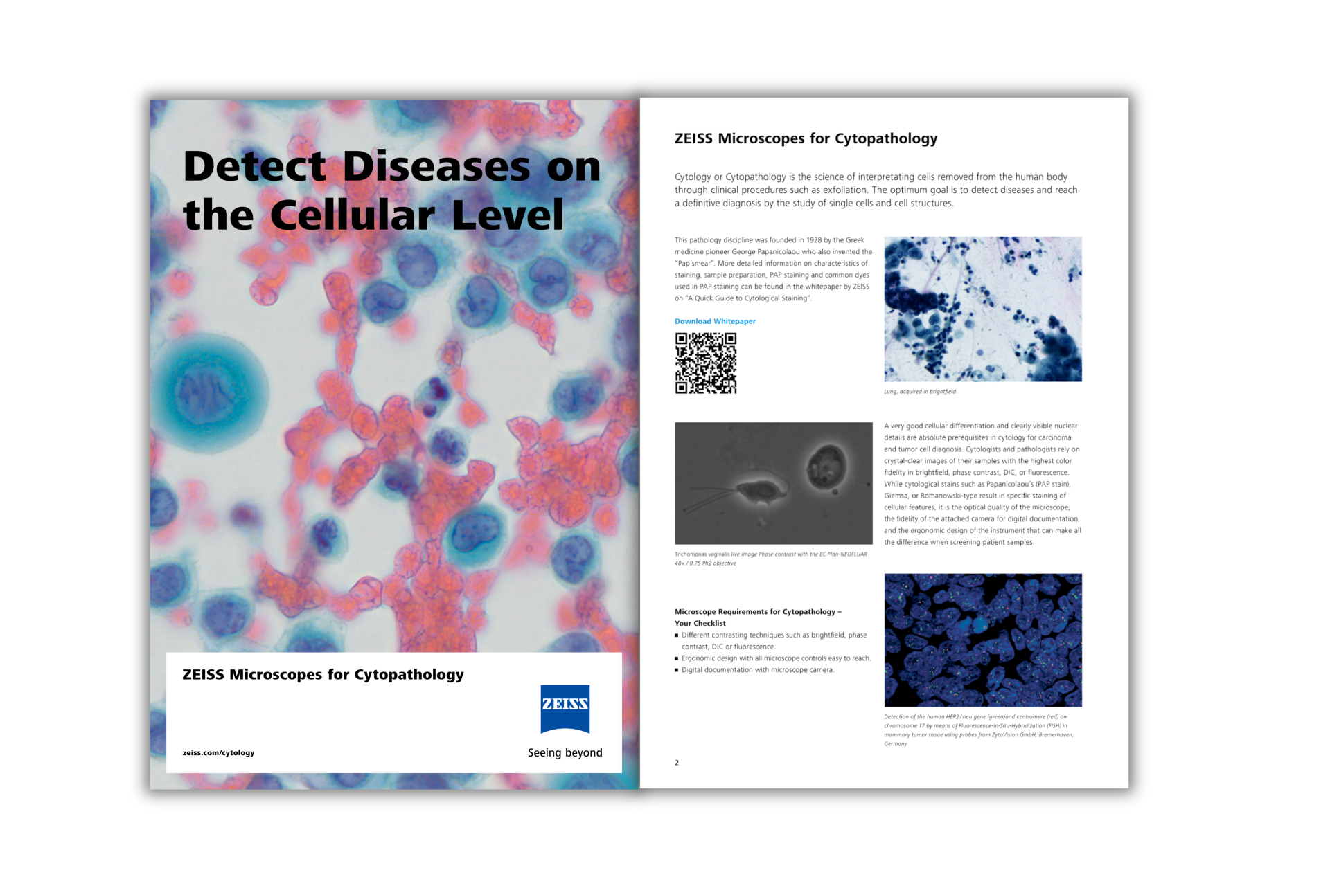
ZEISS 현미경을 활용한 세포병리학에서의 우수성 실현
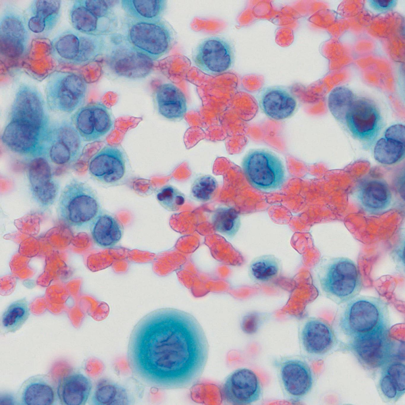
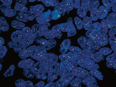
독보적인 디테일의 세포 진단
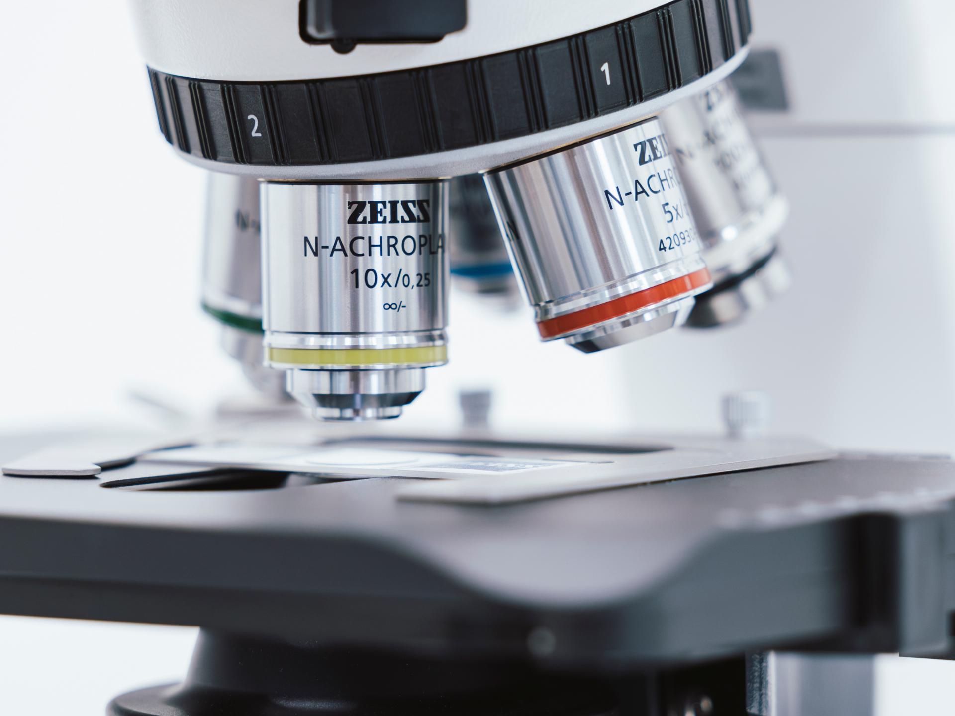
세포 병리학자는 최고 수준의 세포 분화와 정확성이 요구되는 작업을 수행합니다. 세포 병리학에 필요한 요구사항을 고려하여, ZEISS 현미경은 최첨단 기술을 탑재하여 세포 병리학 분석의 복잡성을 충족하도록 설계되었습니다.
![]({"xsmall":"https://www.zeiss.co.kr/content/dam/rms/countries/singapore/apac/cyto-clear1.png/_jcr_content/renditions/original.image_file.100.75.0,0,807,605.file/cyto-clear1.png","small":"https://www.zeiss.co.kr/content/dam/rms/countries/singapore/apac/cyto-clear1.png/_jcr_content/renditions/original.image_file.360.270.0,0,807,605.file/cyto-clear1.png","medium":"https://www.zeiss.co.kr/content/dam/rms/countries/singapore/apac/cyto-clear1.png/_jcr_content/renditions/original.image_file.768.576.0,0,807,605.file/cyto-clear1.png","large":"https://www.zeiss.co.kr/content/dam/rms/countries/singapore/apac/cyto-clear1.png/_jcr_content/renditions/original.image_file.807.605.0,0,807,605.file/cyto-clear1.png","xlarge":"https://www.zeiss.co.kr/content/dam/rms/countries/singapore/apac/cyto-clear1.png/_jcr_content/renditions/original.image_file.807.605.0,0,807,605.file/cyto-clear1.png","xxlarge":"https://www.zeiss.co.kr/content/dam/rms/countries/singapore/apac/cyto-clear1.png/_jcr_content/renditions/original.image_file.807.605.0,0,807,605.file/cyto-clear1.png","max":"https://www.zeiss.co.kr/content/dam/rms/countries/singapore/apac/cyto-clear1.png/_jcr_content/renditions/original.image_file.807.605.0,0,807,605.file/cyto-clear1.png"})
![]({"xsmall":"https://www.zeiss.co.kr/content/dam/rms/countries/singapore/apac/cytofluorescence_her2-centromere_axiocam-mrm_2.jpg/_jcr_content/renditions/original.image_file.100.75.0,0,406,305.file/cytofluorescence_her2-centromere_axiocam-mrm_2.jpg","small":"https://www.zeiss.co.kr/content/dam/rms/countries/singapore/apac/cytofluorescence_her2-centromere_axiocam-mrm_2.jpg/_jcr_content/renditions/original.image_file.360.270.0,0,406,305.file/cytofluorescence_her2-centromere_axiocam-mrm_2.jpg","medium":"https://www.zeiss.co.kr/content/dam/rms/countries/singapore/apac/cytofluorescence_her2-centromere_axiocam-mrm_2.jpg/_jcr_content/renditions/original.image_file.406.305.0,0,406,305.file/cytofluorescence_her2-centromere_axiocam-mrm_2.jpg","large":"https://www.zeiss.co.kr/content/dam/rms/countries/singapore/apac/cytofluorescence_her2-centromere_axiocam-mrm_2.jpg/_jcr_content/renditions/original.image_file.406.305.0,0,406,305.file/cytofluorescence_her2-centromere_axiocam-mrm_2.jpg","xlarge":"https://www.zeiss.co.kr/content/dam/rms/countries/singapore/apac/cytofluorescence_her2-centromere_axiocam-mrm_2.jpg/_jcr_content/renditions/original.image_file.406.305.0,0,406,305.file/cytofluorescence_her2-centromere_axiocam-mrm_2.jpg","xxlarge":"https://www.zeiss.co.kr/content/dam/rms/countries/singapore/apac/cytofluorescence_her2-centromere_axiocam-mrm_2.jpg/_jcr_content/renditions/original.image_file.406.305.0,0,406,305.file/cytofluorescence_her2-centromere_axiocam-mrm_2.jpg","max":"https://www.zeiss.co.kr/content/dam/rms/countries/singapore/apac/cytofluorescence_her2-centromere_axiocam-mrm_2.jpg/_jcr_content/renditions/original.image_file.406.305.0,0,406,305.file/cytofluorescence_her2-centromere_axiocam-mrm_2.jpg"})
![]({"xsmall":"https://www.zeiss.co.kr/content/dam/rms/countries/singapore/apac/cytozeissaxiom-4.jpg/_jcr_content/renditions/original.image_file.100.75.0,1134,3023,3401.file/cytozeissaxiom-4.jpg","small":"https://www.zeiss.co.kr/content/dam/rms/countries/singapore/apac/cytozeissaxiom-4.jpg/_jcr_content/renditions/original.image_file.360.270.0,1134,3023,3401.file/cytozeissaxiom-4.jpg","medium":"https://www.zeiss.co.kr/content/dam/rms/countries/singapore/apac/cytozeissaxiom-4.jpg/_jcr_content/renditions/original.image_file.768.576.0,1134,3023,3401.file/cytozeissaxiom-4.jpg","large":"https://www.zeiss.co.kr/content/dam/rms/countries/singapore/apac/cytozeissaxiom-4.jpg/_jcr_content/renditions/original.image_file.1024.768.0,1134,3023,3401.file/cytozeissaxiom-4.jpg","xlarge":"https://www.zeiss.co.kr/content/dam/rms/countries/singapore/apac/cytozeissaxiom-4.jpg/_jcr_content/renditions/original.image_file.1280.960.0,1134,3023,3401.file/cytozeissaxiom-4.jpg","xxlarge":"https://www.zeiss.co.kr/content/dam/rms/countries/singapore/apac/cytozeissaxiom-4.jpg/_jcr_content/renditions/original.image_file.1440.1080.0,1134,3023,3401.file/cytozeissaxiom-4.jpg","max":"https://www.zeiss.co.kr/content/dam/rms/countries/singapore/apac/cytozeissaxiom-4.jpg/_jcr_content/renditions/original.image_file.1920.1440.0,1134,3023,3401.file/cytozeissaxiom-4.jpg"})
세포 병리학에서의 현미경 실험 과제
- 중요한 분석을 진행해야 하는데 이미징 품질의 한계 때문에 고민이신가요?
- 미세한 세포 해석을 위해 최신 염색 솔루션이 필요하신가요?
- 인체공학적 불편함 때문에 집중력과 생산성이 떨어지고 있으신가요?
- 보다 효율적인 워크플로우 디지털 통합을 찾고 계신가요?
Insights Package를 통해
- PAP 및 로마노프스키 염색을 포함한 세포 병리학에 적합한 염색의 특성에 대해 확인하세요.
- 도말, 고정, 투과화, 슬라이드 장착과 같은 세포 염색을 위해 샘플을 준비하는 방법과 단계를 알아보세요.
- 일반적으로 사용되는 염색, 시료 준비 및 염색 원리를 포함한 PAP 염색의 원리에 대해 파악하세요.
- 세포 병리 분석의 복잡한 요구 사항을 충족하는 ZEISS 현미경 솔루션에 대해 알아보세요.
Cytology Insights Package에서 확인하세요!
- 세포 염색 가이드
- Quick Guide: 세포 병리학을 위한 ZEISS 현미경